
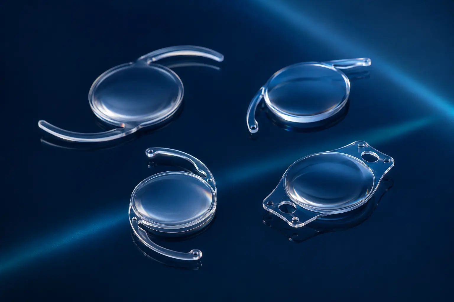
What an ICL is—and is not
An ICL is positioned within the eye, typically behind the iris and in front of the crystalline lens. It works with the existing optics rather than reshaping the cornea. Toric models can also correct suitable astigmatism.
It is not the same as the IOL used after cataract removal, because the natural lens stays in place. It is also not a contact lens and cannot be inserted or removed without surgery.
Candidacy and essential measurements
The prescription should be stable and the anterior chamber must have adequate dimensions for the chosen lens. Endothelial cell density, angle anatomy, pupil, natural-lens clarity and retinal status are assessed.
Sizing is critical. Measurements help predict vault—the space between ICL and natural lens. Too little or too much vault can increase cataract, angle or pressure concerns.
What happens during surgery
Through a small corneal incision, the folded lens is placed inside the eye and positioned behind the iris. Anaesthesia, medication and whether both eyes are treated together depend on the plan and setting.
The operation may be brief, but it should not be marketed as trivial. Sterility, lens orientation, complete removal of viscoelastic where used and early pressure monitoring are important.
Early recovery
Vision may improve quickly, with temporary blur, glare, mild discomfort or light sensitivity. Drops are used to control inflammation and reduce infection risk. The eye must be protected from rubbing, contamination and unapproved activity.
Early review checks vision, pressure, inflammation, lens position and vault. Severe pain, nausea with eye pain, increasing redness or sudden loss of vision require urgent assessment.
Long-term risks and monitoring
Potential issues include cataract, endothelial cell loss, pressure rise, pigment dispersion, lens rotation or decentration, incorrect vault, infection and retinal events related to high myopia. Some problems may require repositioning, exchange or removal.
Long-term examinations monitor the cornea, pressure, vault, natural lens and retina. Good early vision is not a reason to stop follow-up.
How ICL compares with corneal laser
ICL preserves corneal tissue and may offer excellent optical quality in high corrections. Corneal laser avoids entering the eye and does not leave an implant, but it changes corneal structure and can worsen dry-eye symptoms.
The right comparison is individual: tissue calculations versus internal anatomy, ocular-surface status, prescription magnitude, age and willingness for long-term lens monitoring.
Useful questions for your consultation
Use these prompts to keep the discussion connected to your measurements, risks and visual goals.
- What is my endothelial cell count?
- How was lens size selected?
- What vault range are you aiming for?
- What long-term follow-up schedule do you recommend?
Frequently asked questions
Will I feel the ICL inside my eye?
Normally it is not felt like a contact lens because it is inside the eye, but discomfort or pressure symptoms after surgery require review.
Does ICL cause cataract?
Cataract is a recognized risk, influenced by age, vault, lens model and other factors. Monitoring the natural lens is part of follow-up.
Can ICL correct astigmatism?
Toric ICL models can correct suitable astigmatism when sizing and rotational planning are appropriate.
Is follow-up lifelong?
Regular long-term eye examinations are advisable to monitor pressure, endothelium, vault, natural lens and retina.
Medical sources and further reading
This article was reviewed against the following patient-information resources. Individual recommendations still depend on examination.
- U.S. FDA — Phakic intraocular lens risks
- National Eye Institute — Cataracts and intraocular lenses
- American Academy of Ophthalmology — Cataract surgery
- Alcon — Cataract lens portfolio
- Johnson & Johnson Vision — TECNIS Eyhance
- Johnson & Johnson Vision — TECNIS PureSee
- Bausch + Lomb Surgical — Cataract portfolio