
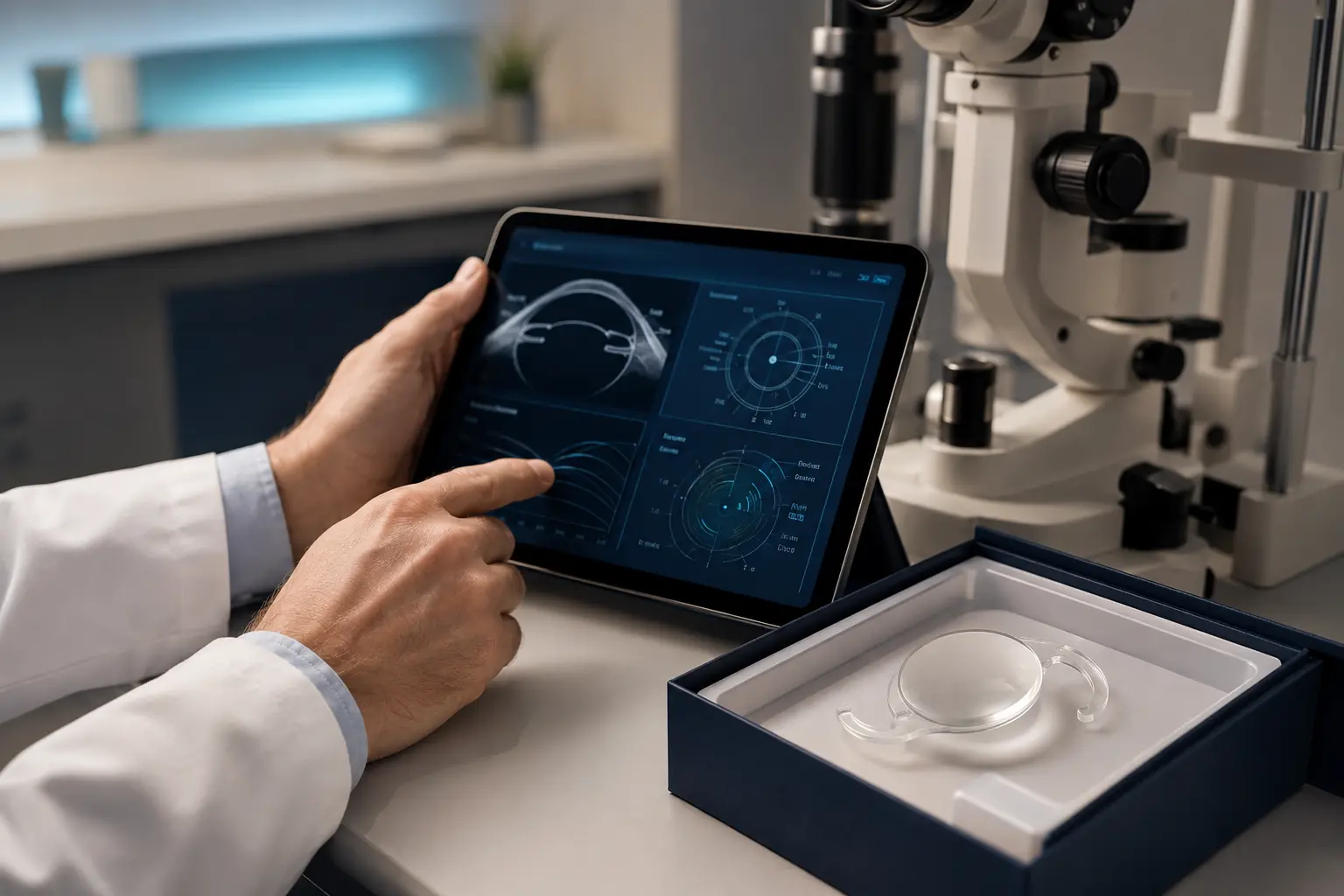
Preoperative assessment and biometry
The surgeon confirms that cataract is a meaningful cause of the visual problem and evaluates the cornea, anterior chamber, pupil, pressure, optic nerve and retina. OCT or other tests may be needed when macular disease is suspected.
Biometry measures axial length and corneal power to calculate the IOL. Ocular-surface disease can distort keratometry, so dry eye or lid disease may need treatment before final measurements.
Choosing the target and lens
IOL power is chosen for a target such as distance, near, mild monovision or a broader range with a presbyopia-correcting design. Toric correction may be considered for regular corneal astigmatism.
The decision should include driving, reading, computer distance, previous refractive surgery, ocular disease and tolerance for halos. No formula or lens guarantees an exact glasses-free result.
Anaesthesia and preparation
Most routine surgery is performed with topical or local anaesthesia, with sedation according to the setting and patient. The eye is cleaned, draped and held open gently.
Patients are usually awake and may see light or movement without seeing surgical detail. Medication instructions—especially anticoagulants, diabetes treatments and alpha-blockers—must be individualized rather than stopped without medical advice.
Phacoemulsification and IOL placement
Through small incisions, the surgeon creates an opening in the front capsule. Ultrasound phacoemulsification divides and removes the cataract while preserving the capsular bag when possible.
Cortical material is cleaned, a folded IOL is inserted and opened within the bag, and the incisions usually seal without stitches. Complex cataracts may need pupil devices, capsular support, sutures or a different lens position.
Immediately after surgery
Vision may be blurred from drops, corneal swelling or the healing response. A shield may be advised, and antibiotic or anti-inflammatory drops are prescribed according to the surgeon’s plan.
The first review checks pressure, wound, cornea, inflammation and lens position. Pain that is severe or increasing, nausea with eye pain, marked redness or sudden vision loss requires urgent contact.
The second eye and final glasses
Timing of the other eye depends on local practice, recovery, anisometropia and medical considerations. Final refraction is performed after sufficient stability, but the exact time varies.
If a residual prescription remains, options may include glasses, contact lenses or selected additional procedures after the eye stabilizes. The first step is identifying whether blur comes from refraction, surface, retina, capsule or another cause.
Useful questions for your consultation
Use these prompts to keep the discussion connected to your measurements, risks and visual goals.
- What limits my expected vision?
- Which IOL target fits my daily life?
- Could previous LASIK affect the calculation?
- What urgent symptoms should I report?
Frequently asked questions
Is cataract surgery painful?
Numbing anaesthesia makes routine surgery comfortable for most patients, though pressure, light or mild discomfort may be noticed.
Is laser always used?
Standard phaco uses ultrasound. A femtosecond laser may perform selected steps in some settings, but it is not required for excellent surgery.
Are stitches always needed?
Small incisions commonly seal without stitches; complex or less stable wounds may need one.
Can both eyes be treated together?
Practice varies. Benefits and bilateral risks must be considered individually.
Medical sources and further reading
This article was reviewed against the following patient-information resources. Individual recommendations still depend on examination.